
In 2003, a German scientist named Heiko Braak made a wild hypothesis about the nature of Parkinson’s disease.1 He published a paper suggesting that environmental pathogens cause Parkinson’s disease by entering the stomach and traveling to the brain through connections. At the time, the evidence to support this was two-fold. There was the argument that the pathway of neurons dying in the brain suggested the source of the problem started outside the brain, and the identification of alpha-synuclein (a-syn) aggregates, a component of Parkinson’s disease, in the neurons surrounding the gut.2 Finally, in 2019, this study provides definitive evidence supporting Braak’s hypothesis.
Three takeaways to tell your friends:
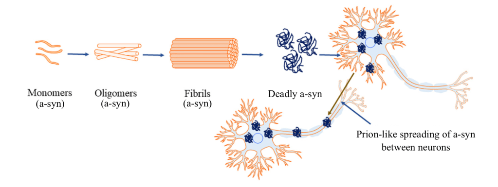
- Alpha-synuclein (a-syn) travels between neurons, sticks together to form aggregates, and leads to neuron death in Parkinson’s disease.
- A-syn delivered to the small intestine leads to a-syn accumulation in the brain, loss of Dopamine neurons, lower dopamine levels, and Parkinson’s-related behaviors.3
- A-syn delivered to the small intestine does not create Parkinson’s symptoms when the stomach-to-brain connection is cut.3
The dopamine killer, Parkinson’s disease; if you are unfamiliar, the neurons that produce one of your essential neurotransmitters, Dopamine, die. If you are familiar with Parkinson’s disease, did you know that it falls under the net of diseases known as synucleinopathies (pronounced sin-nu-clee-in-ah-pa-thees)?4 The name comes from the protein known to cause the diseases, alpha-synuclein (a-syn). Typically involved in neurotransmitter release, the a-syn protein can misfold, creating a catastrophe for neurons (Figure 1). Protein folding is an essential process for healthy protein function. Additionally, a-syn may act similarly to prions.5 Prions are misfolded forms of a protein that convert other healthy forms of that protein into the misfolded form, spreading its diseased, misfolded form, rapidly. So, these a-syn proteins travel between neurons, making the healthy a-syn proteins misfold, stick together, and form deadly aggregates? (Figure 1) Yes, that’s synucleinopathies, aka. Parkinson’s disease, but where do they originate?
As stated, there was evidence in 2003 of aggregated a-syn in the neurons adjacent to the intestine2; in 2014, a study of fifteen deceased patients with synucleinopathies found thirteen had a-syn aggregates in both the gastrointestinal tract and the stomach-to-brain nerve connection, known as the vagus nerve,7 providing more support to Braak’s hypothesis.
With this in mind, the researchers sought to solve it once and for all. To achieve this, they delivered misfolded a-syn to the small intestines of mice, with or without cutting the gut’s connection to the brain, the vagus nerve, to see if Parkinson’s disease symptoms progress to the brain. Oh boy, they tested everything.
Within three months of a-syn injection, with an intact vagus nerve, a-syn travels to the brain, specifically to the brain region most impacted by Parkinson’s disease (Substantia Nigra). At seven months, there is a significant loss of dopamine neurons and dopamine levels in the brain.3
In comparison, seven months after a-syn injection, with a cut vagus nerve, no a-syn was detected in the mouse brains.3 Additionally, there was no change in dopamine neurons or dopamine levels in the brain (Figure 2).

On to the plethora of behavioral tests. Here is the purpose of each test: spatial learning and memory, recognition memory, brain-region-specific memory, short-term working memory, fine-motor skills, balance, grip strength, anxiety, locomotion and anxiety, depression, and smell ability. Let’s pour one out for the poor grad students who ran these behavioral tests.
Now, the a-syn injected mice with an intact vagus nerve have Parkinson’s pathology, and these tests highlight that. I am not joking when I say the vagus nerve mice performed significantly worse on every task.3 Guess which mice performed identically to healthy control mice on every task? The a-syn-injected mice with a cut vagus nerve.3 Therefore, Parkinson’s disease pathology can start in the intestines and spread to the brain. This evidence supports Braak’s hypothesis and pushes it to the forefront of Parkinson’s research.
There were other papers simultaneously publishing mildly contradictory results. So, in their discussion section, they compare their methods with the other papers’ and provide sources for why they believe results occurred. Magnificent.
REFERENCES
1. Braak H, Del Tredici K, Rub U, de Vos RA, Jansen Steur EN, Braak E. Staging of brain pathology related to sporadic Parkinson’s disease. Neurobiol Aging. 2003;24(2):197-211.
2. Braak H, Rub U, Gai WP, Del Tredici K. Idiopathic Parkinson’s disease: possible routes by which vulnerable neuronal types may be subject to neuroinvasion by an unknown pathogen. J Neural Transm (Vienna). 2003;110(5):517-36.
3. Kim S, Kwon SH, Kam TI, Panicker N, Karuppagounder SS, Lee S, et al. Transneuronal Propagation of Pathologic alpha-Synuclein from the Gut to the Brain Models Parkinson’s Disease. Neuron. 2019;103(4):627-41 e7.
4. Koga S, Sekiya H, Kondru N, Ross OA, Dickson DW. Neuropathology and molecular diagnosis of Synucleinopathies. Mol Neurodegener. 2021;16(1):83.
5. Luk KC, Kehm V, Carroll J, Zhang B, O’Brien P, Trojanowski JQ, et al. Pathological alpha-synuclein transmission initiates Parkinson-like neurodegeneration in nontransgenic mice. Science. 2012;338(6109):949-53.
6. Bohra SK, Achar RR, Chidambaram SB, Pellegrino C, Laurin J, Masoodi M, et al. Current perspectives on mitochondrial dysfunction in migraine. Eur J Neurosci. 2022;56(1):3738-54.
7. Gelpi E, Navarro-Otano J, Tolosa E, Gaig C, Compta Y, Rey MJ, et al. Multiple organ involvement by alpha-synuclein pathology in Lewy body disorders. Mov Disord. 2014;29(8):1010-8.

Leave a comment